
Compare muscle and nervous tissue without confusing fields and populations
Muscle tissue generates force through organized contractile machinery; nervous tissue receives, integrates and transmits signals. Both can be electrically excitable, so excitability alone does not identify one tissue class. Skeletal muscle typically has long striated multinucleate fibers, cardiac muscle has striated interconnected cells with central nuclei and specialized junctions, and smooth muscle has spindle-shaped cells without visible cross-striations. Involuntary regulation does not mean a tissue lacks electrical or chemical control.
Learning outcome: Compare the actual muscle and neuron images and retain two discriminating visible features plus one unresolved feature. Then compare areas 20/80 versus 50/50 in the synthetic packet, preserving tissue fractions and total area.
Starting recap
Use subject-relative anatomical directions, actual licensed tissue images, compartment conservation and explicit feedback/system boundaries; distinguish structural evidence from functional and clinical claims.
All lessons are open directly. Your saved experiments remain when you leave and return.
Try
Field A has area 20 and target fraction 0.8; field B has area 80 and target fraction 0.2. Target areas are 16 and 16, so the total fraction is 32/100=0.32. The equal-field mean is 0.5 and answers a different question.
Open the experimentPredict
Can a small selected field make an equally weighted average overstate the fraction across all sampled tissue?
Experiment
Compare the actual muscle and neuron images and retain two discriminating visible features plus one unresolved feature. Then compare areas 20/80 versus 50/50 in the synthetic packet, preserving tissue fractions and total area.
Weight tissue evidence by sampled area
Worked teaching model · synthetic values · Original segmented teaching fields have total area 100; field B target fraction 0.2. Compare area-weighted fraction with equally weighting two fields. Counts are not derived from the supplied micrographs and do not estimate whole-organ cell numbers.
Reset keeps your prediction, captures and practice history.
A field average and a tissue-area fraction have different denominators.
Neither fraction automatically counts cells or represents the whole organ. Selecting attractive fields, different section thicknesses and repeated fields from one specimen can bias inference or create pseudoreplication.
| Quantity | Value | Unit |
|---|---|---|
| Field A target area | 16 | area units |
| Field B target area | 16 | area units |
| Area-weighted fraction | 0.3200000 | fraction |
| Equal-field mean | 0.5000000 | fraction |
Original bounded models, not measurements of the accompanying actual micrographs or any person. Retain settings, predictions and limitations in the notebook.
A field average and a tissue-area fraction have different denominators.
Neither fraction automatically counts cells or represents the whole organ. Selecting attractive fields, different section thicknesses and repeated fields from one specimen can bias inference or create pseudoreplication.
Captures preserve recipes and replay their calculated results. They are synthetic model evidence, not physical measurements.
Understand
1. Define the structure and boundary
Muscle tissue generates force through organized contractile machinery; nervous tissue receives, integrates and transmits signals. Both can be electrically excitable, so excitability alone does not identify one tissue class. Skeletal muscle typically has long striated multinucleate fibers, cardiac muscle has striated interconnected cells with central nuclei and specialized junctions, and smooth muscle has spindle-shaped cells without visible cross-striations. Involuntary regulation does not mean a tissue lacks electrical or chemical control.
2. Connect anatomical evidence and mechanism
The actual CC0 cardiac image shows elongated pink fibers with fine transverse striations and nuclei along the fibers. Use source identity plus multiple visible features, not one dark line, to support the label. Intercalated discs provide mechanical attachment and electrical coupling, but their molecular components are not resolved here; do not claim to have measured conduction from the image. The separate actual motor-neuron image shows a large cell body with branching processes amid many smaller stained nuclei.
Neurons have a soma containing the nucleus and processes specialized for receiving and transmitting signals. Glial cells contribute support, homeostasis, insulation and immune surveillance; they are not merely passive filler. A single light micrograph cannot reliably assign every visible process as axon versus dendrite or every small nucleus to a specific glial subtype. Retain such uncertainty rather than drawing a definitive circuit from one section. No live stimulation, personal testing or specimen handling is requested.
The numerical action uses a separate original segmented-field packet, not pixel measurements of these images. Field A occupies a fraction of the sampled area and contains a supplied target-tissue fraction; field B occupies the rest. Total target area divided by total sampled area is an area-weighted fraction. Averaging the two fractions equally estimates an equal-field quantity instead. Neither automatically estimates cell number, organ-wide volume or independent biological replication.
Observe actual tissue images before accepting a label
These are actual published micrographs, not generated images or the numerical model’s invented measurements. Retain two visible features, a tentative tissue class and one unresolved feature in your lesson notebook. The accessible descriptions supply equivalent observations; using them or the public source label is guided evidence, not a blinded identification test.
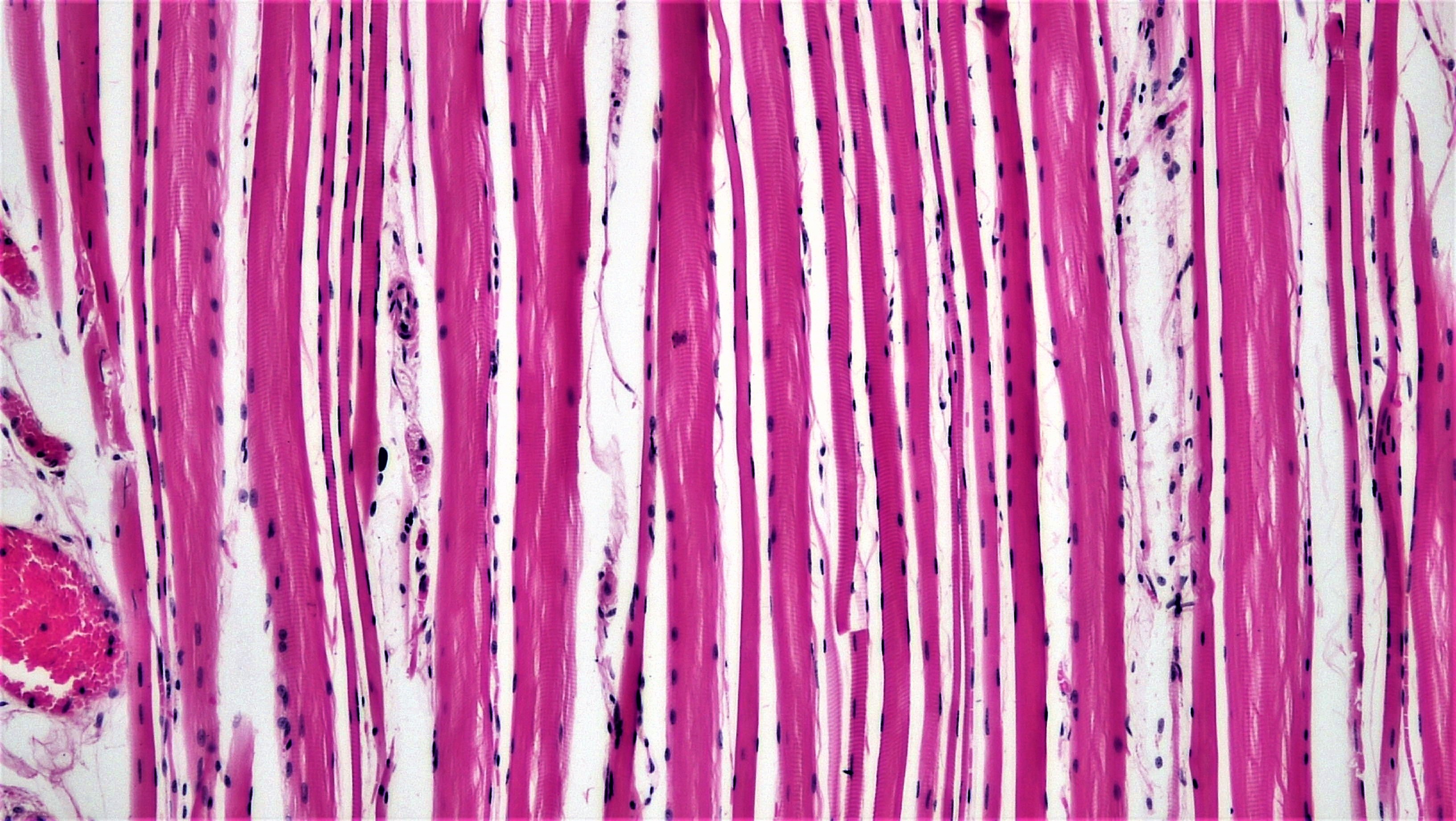
Compare source identity, visible evidence and limits
The source labels cardiac muscle. Multiple features plus provenance support this guided identification; image geometry alone does not demonstrate contraction, electrical coupling or organ performance. Skeletal and cardiac muscle both have striations, so that single feature is insufficient.
Berkshire Community College Bioscience Image Library; 5 April 2018. Source and file history · CC0 1.0 dedication · Download unchanged image. Checked 30 October 2026. No endorsement or diagnostic use is implied.

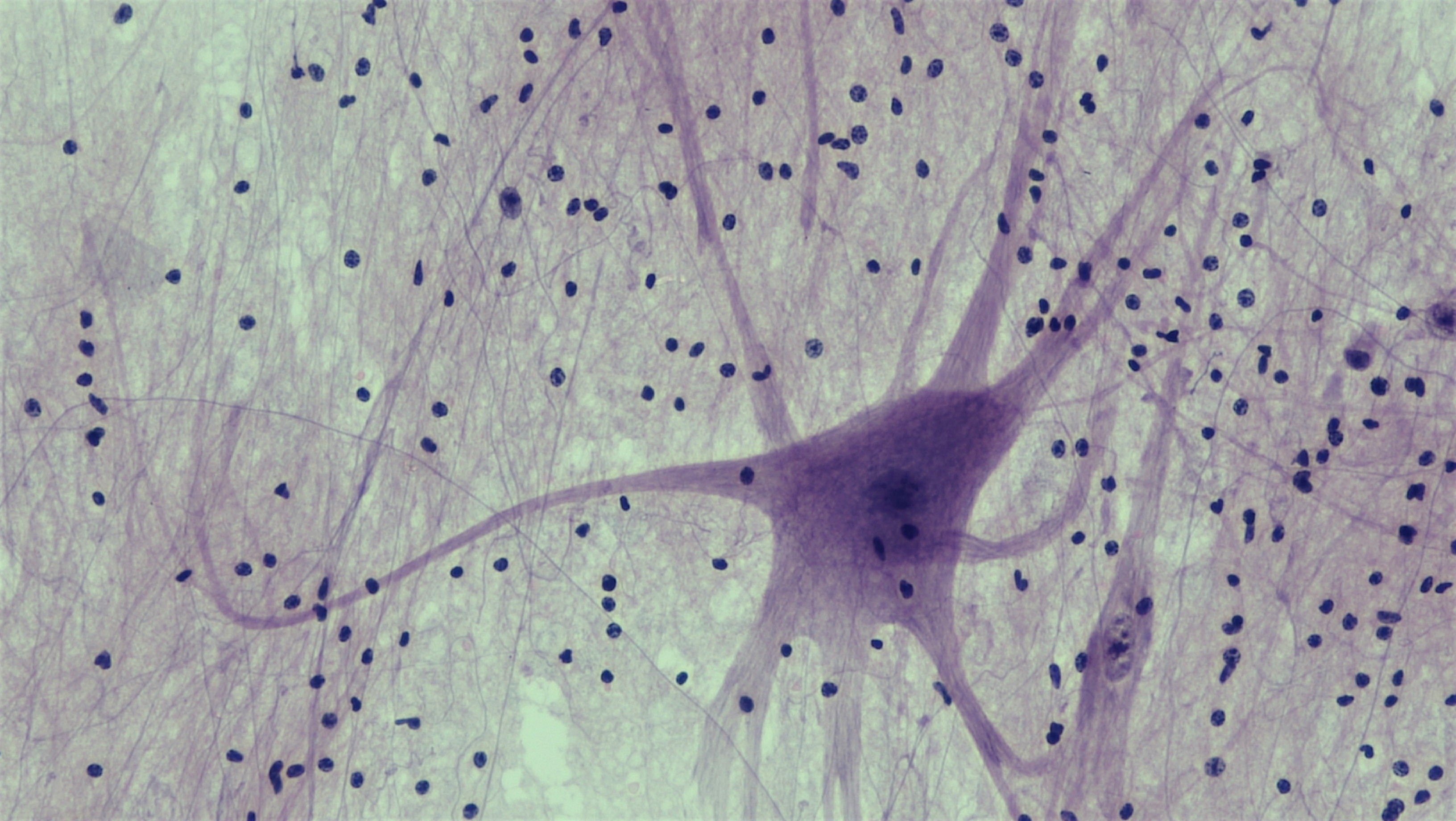
Compare source identity, visible evidence and limits
The source identifies a spinal-cord motor neuron preparation. A large soma with multiple processes supports neural architecture, but this field does not resolve every axon/dendrite identity, small-cell subtype or direction of functional signaling.
Berkshire Community College Bioscience Image Library; 3 May 2018. Source and file history · CC0 1.0 dedication · Download unchanged image. Checked 30 October 2026. No endorsement or diagnostic use is implied.

Download exact source, license, dimensions and SHA-256 provenance. For independent practical evidence, a qualified reviewer must supply a genuinely unfamiliar licensed specimen/image and assess identification, uncertainty and reasoning; viewing these labeled examples does not fulfill that gate.
3. Audit an original worked comparison
Field A has area 20 and target fraction 0.8; field B has area 80 and target fraction 0.2. Target areas are 16 and 16, so the total fraction is 32/100=0.32. The equal-field mean is 0.5 and answers a different question.
4. Predict and investigate
Predict first: Can a small selected field make an equally weighted average overstate the fraction across all sampled tissue?
Compare the actual muscle and neuron images and retain two discriminating visible features plus one unresolved feature. Then compare areas 20/80 versus 50/50 in the synthetic packet, preserving tissue fractions and total area.
5. Transfer to a changed case
Apply the mechanism to a changed boundary
Field A area 60 has target fraction 0.6; field B area 40 has target fraction 0.2. Find the area-weighted fraction and equal-field mean.
Retain the first response, units, uncertainty and any prior feedback exposure in the notebook before comparing this public explanation.
Compare a worked response
Target area is 36+8=44 of 100, so the weighted fraction is 0.44. The equal-field mean is 0.4. Neither is a whole-organ cell count.
6. Retain an evidence-based defense
Use the Anatomy and Physiology course workspace to preserve original reasoning, actual image observations with uncertainty, model settings and a reviewer-supplied new transfer. Public feedback is guided practice, not independent mastery or clinical assessment. No examination, personal health data, specimen collection or physiological challenge is requested.
Supporting anatomical mechanism, checked 30 October 2026. Numerical packets and diagrams are original teaching models; actual tissue images have separate attribution, download and CC0 provenance records.
Apply
Public practice checks a specific claim. It does not establish independent mastery.
Field A area 60 has target fraction 0.6; field B area 40 has target fraction 0.2. Find the area-weighted fraction and equal-field mean.
Continue
Completion requires a saved prediction, at least one capture and a reflection. Presence checks do not grade your reasoning.
Skipping retains experiments and awards no mastery.
Save a science backup or import one
Science work is stored locally in this browser, separately from mathematics and AI. It does not sync automatically. Use this science-only backup; this checkout's older all-subject export does not include science.
A legacy file is available only when the complete snapshot fits the 36 MB limit. For larger snapshots, prepare a complete file set below; never discard notebooks to make a smaller file.
Matching imported notebook drafts replace their current drafts. Omitted lessons and known first attempts remain. Export your current work first if you want to retain both drafts.
Complete file sets and course backups
A file set can hold more work than one science file. Keep its manifest and every part together. Each file is limited to 36 MB. This format needs this version of LearnAI or a compatible newer version; older versions can use only the legacy single-file backup above.
The set includes every saved science course and global continuation at this moment. Later edits need a new export. A conflicting tab exports its own visible work, which may differ from saved work in another tab.
0 file(s) selected. File order and filenames do not determine identity. Missing, changed or conflicting parts block the entire restore; no good part is applied by itself.
Science storage: loading. No account or remote computation is required.