
Connect extracellular matrix architecture to directional support
Connective tissues contain cells within an extracellular matrix whose ground substance and fibers strongly shape function. Fibroblasts maintain many connective matrices; adipocytes store lipid; cartilage and bone have specialized cells and matrices; blood has a fluid matrix. These are not simply gaps between more important tissues. Matrix composition, hydration, mineralization and organization can support, cushion, transport or transmit forces in different contexts.
Learning outcome: Compare θ=0°,45°,90° at f=0.5, then change f at fixed 45°. Retain the two projection steps and identify features in the actual tissue image that the single-direction model omits.
Starting recap
Use subject-relative anatomical directions, actual licensed tissue images, compartment conservation and explicit feedback/system boundaries; distinguish structural evidence from functional and clinical claims.
All lessons are open directly. Your saved experiments remain when you leave and return.
Try
At f=0.5 and θ=45°, cos⁴θ=1/4. The projected fiber contribution is 9×0.5×0.25=1.125 and total K=2.125. At θ=90° the stipulated axial fiber contribution is zero, but the matrix baseline remains 1.
Open the experimentPredict
Must equal fiber fractions yield equal stiffness when the load direction changes?
Experiment
Compare θ=0°,45°,90° at f=0.5, then change f at fixed 45°. Retain the two projection steps and identify features in the actual tissue image that the single-direction model omits.
Test an explicit matrix-fiber orientation model
Worked teaching model · synthetic values · Small imposed uniaxial strain, no transverse strain or rotation: axial fiber strain projects by cos² θ and axial stress contribution by another cos² θ. Supplied normalized stiffness K=1+9f cos⁴ θ is a toy composite, not measured tissue stiffness.
Reset keeps your prediction, captures and practice history.
The same fiber amount can contribute differently along different loading axes.
The cos⁴ rule follows the explicitly fixed deformation assumptions. It is not a fit to the elastic-tissue image; actual matrix chemistry, crimp, remodeling and multidirectional fibers require independent measurements.
| Quantity | Value | Unit |
|---|---|---|
| Matrix baseline contribution | 1 | relative stiffness |
| Projected fiber contribution | 1.125000 | relative stiffness |
| Total normalized stiffness | 2.125000 | relative stiffness |
Original bounded models, not measurements of the accompanying actual micrographs or any person. Retain settings, predictions and limitations in the notebook.
The same fiber amount can contribute differently along different loading axes.
The cos⁴ rule follows the explicitly fixed deformation assumptions. It is not a fit to the elastic-tissue image; actual matrix chemistry, crimp, remodeling and multidirectional fibers require independent measurements.
Captures preserve recipes and replay their calculated results. They are synthetic model evidence, not physical measurements.
Understand
1. Define the structure and boundary
Connective tissues contain cells within an extracellular matrix whose ground substance and fibers strongly shape function. Fibroblasts maintain many connective matrices; adipocytes store lipid; cartilage and bone have specialized cells and matrices; blood has a fluid matrix. These are not simply gaps between more important tissues. Matrix composition, hydration, mineralization and organization can support, cushion, transport or transmit forces in different contexts.
2. Connect anatomical evidence and mechanism
Collagen fibers resist tensile loading, elastic fibers can stretch and recoil, and reticular networks provide fine supporting frameworks. Dense regular connective arrangements favor a dominant loading direction, whereas multidirectional arrangements distribute support across directions. A dark line in a particular stain is not by itself a mechanical test. The actual CC0 elastic-tissue image contains dark branching fibers among thicker pink bundles and scattered nuclei; record that visible organization separately from the source’s tissue label.
The model is an explicit small-strain composite, not an estimate from the image. A fraction f of aligned load-bearing fibers lies at angle θ to a prescribed uniaxial deformation. With transverse strain and rotation suppressed, fiber axial strain scales as cos² θ; projecting its stress back onto the loading axis supplies another cos² factor. We stipulate normalized stiffness K=1+9f cos⁴ θ, including a constant matrix baseline of 1. The coefficient 9 is a teaching choice, not a material constant.
Real connective tissues can straighten crimped fibers, rotate, change hydration and remodel, so their force–deformation curves need not follow this linear expression. Compare direction at fixed composition before attributing a stiffness difference to changed protein amount. A paired directional measurement from one sample is not independent evidence from many specimens; sample identity, loading protocol and preparation must be retained in any practical review.
Observe actual tissue images before accepting a label
These are actual published micrographs, not generated images or the numerical model’s invented measurements. Retain two visible features, a tentative tissue class and one unresolved feature in your lesson notebook. The accessible descriptions supply equivalent observations; using them or the public source label is guided evidence, not a blinded identification test.
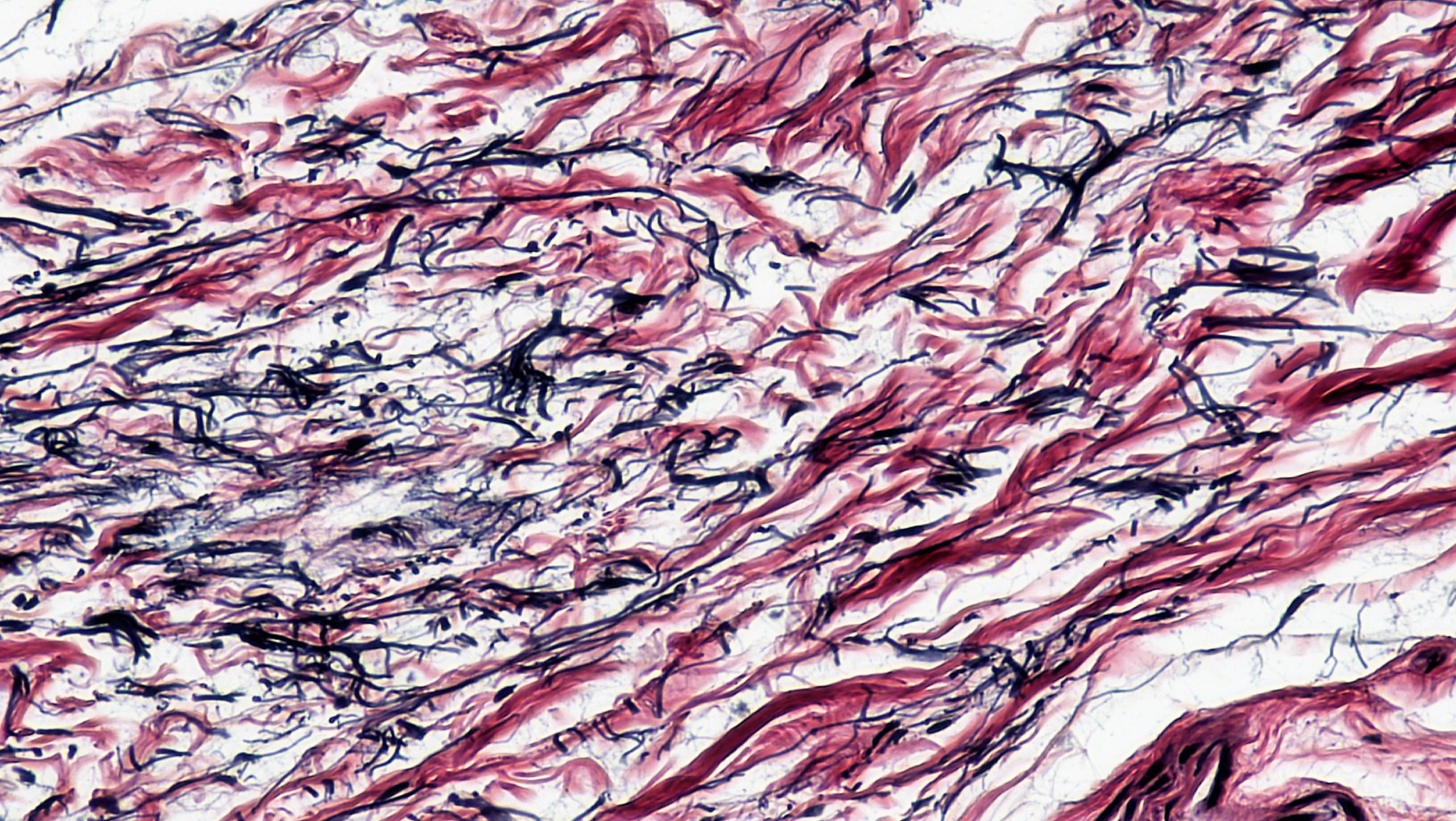
Compare source identity, visible evidence and limits
The source identifies human elastic connective tissue. The visible network is consistent with extracellular fiber organization; exact fiber chemistry and mechanical properties cannot be uniquely inferred from color alone. The single-angle model is deliberately not fitted to this specimen.
Berkshire Community College Bioscience Image Library; 26 March 2018. Source and file history · CC0 1.0 dedication · Download unchanged image. Checked 30 October 2026. No endorsement or diagnostic use is implied.

Download exact source, license, dimensions and SHA-256 provenance. For independent practical evidence, a qualified reviewer must supply a genuinely unfamiliar licensed specimen/image and assess identification, uncertainty and reasoning; viewing these labeled examples does not fulfill that gate.
3. Audit an original worked comparison
At f=0.5 and θ=45°, cos⁴θ=1/4. The projected fiber contribution is 9×0.5×0.25=1.125 and total K=2.125. At θ=90° the stipulated axial fiber contribution is zero, but the matrix baseline remains 1.
4. Predict and investigate
Predict first: Must equal fiber fractions yield equal stiffness when the load direction changes?
Compare θ=0°,45°,90° at f=0.5, then change f at fixed 45°. Retain the two projection steps and identify features in the actual tissue image that the single-direction model omits.
5. Transfer to a changed case
Apply the mechanism to a changed boundary
For f=1 and θ=60°, compute K. Does this establish the stiffness of the supplied elastic-tissue specimen?
Retain the first response, units, uncertainty and any prior feedback exposure in the notebook before comparing this public explanation.
Compare a worked response
cos60°=1/2, so K=1+9/16=1.5625. No: fiber fraction, constitutive response and deformation constraints were supplied, not measured from that specimen.
6. Retain an evidence-based defense
Use the Anatomy and Physiology course workspace to preserve original reasoning, actual image observations with uncertainty, model settings and a reviewer-supplied new transfer. Public feedback is guided practice, not independent mastery or clinical assessment. No examination, personal health data, specimen collection or physiological challenge is requested.
Supporting anatomical mechanism, checked 30 October 2026. Numerical packets and diagrams are original teaching models; actual tissue images have separate attribution, download and CC0 provenance records.
Apply
Public practice checks a specific claim. It does not establish independent mastery.
For f=1 and θ=60°, compute K. Does this establish the stiffness of the supplied elastic-tissue specimen?
Continue
Completion requires a saved prediction, at least one capture and a reflection. Presence checks do not grade your reasoning.
Skipping retains experiments and awards no mastery.
Save a science backup or import one
Science work is stored locally in this browser, separately from mathematics and AI. It does not sync automatically. Use this science-only backup; this checkout's older all-subject export does not include science.
A legacy file is available only when the complete snapshot fits the 36 MB limit. For larger snapshots, prepare a complete file set below; never discard notebooks to make a smaller file.
Matching imported notebook drafts replace their current drafts. Omitted lessons and known first attempts remain. Export your current work first if you want to retain both drafts.
Complete file sets and course backups
A file set can hold more work than one science file. Keep its manifest and every part together. Each file is limited to 36 MB. This format needs this version of LearnAI or a compatible newer version; older versions can use only the legacy single-file backup above.
The set includes every saved science course and global continuation at this moment. Later edits need a new export. A conflicting tab exports its own visible work, which may differ from saved work in another tab.
0 file(s) selected. File order and filenames do not determine identity. Missing, changed or conflicting parts block the entire restore; no good part is applied by itself.
Science storage: loading. No account or remote computation is required.