
Audit actual micrographs before counting or identifying objects
Two actual public-domain images accompany this lesson: a DIC image of yeast attributed to Masur, with a later rotation credited to Andrew Pertsev, and a digital-microscope field attributed to Cucumberkvp and labeled Paramecium bursaria by its creator. Exact source pages, authorship, reuse declarations, original bytes and hashes are retained. Those source labels are provenance, not a blinded identification achieved by the learner.
Learning outcome: Inspect both actual images and retain two visible features, one unresolved claim and exact source identity. Then compare both counting rules on the separate original schematic, including and excluding the ambiguous feature. Explain why neither actual species nor viable count follows from that schematic.
Starting recap
Compare microbial structures and imaging evidence with calibrated geometry, explicit detection and object-definition limits, actual source provenance and no culture or diagnostic inference.
All lessons are open directly. Your saved experiments remain when you leave and return.
Try
The original schematic has 5 unambiguous connected objects but 7 unambiguous bodies. Including the faint feature gives 6 or 8 depending on the declared counting rule. Neither answer establishes viability.
Open the experimentPredict
Can two defensible counting rules give different totals for the same attached objects?
Experiment
Inspect both actual images and retain two visible features, one unresolved claim and exact source identity. Then compare both counting rules on the separate original schematic, including and excluding the ambiguous feature. Explain why neither actual species nor viable count follows from that schematic.
Count visible objects without declaring viable cells
Worked teaching model · synthetic values · Original schematic field, not quantitative segmentation of the actual yeast image: three isolated oval bodies, two attached pairs (four bodies) and one ambiguous faint circular artifact. Connected-object rule counts5+b; body rule7+b. A still image does not establish viability, species or cell division rate.
Reset keeps your prediction, captures and practice history.
A reported count depends on the object definition and treatment of uncertain features.
The original diagram is separate from the actual micrographs. Attached bodies may be compatible with budding, but a single field cannot establish a division rate, species, viability or prevalence.
| Quantity | Value | Unit |
|---|---|---|
| Unambiguous connected objects | 5 | objects |
| Resolved unambiguous oval bodies | 7 | bodies |
| Chosen reported count | 5 | objects under selected rule |
| Ambiguous feature included | 0 | 0 no / 1 yes |
Original diagrams and supplied measurements are not actual microscopy data. The separately labeled public-domain images retain their own source and measurement limits. No biological material is handled.
A reported count depends on the object definition and treatment of uncertain features.
The original diagram is separate from the actual micrographs. Attached bodies may be compatible with budding, but a single field cannot establish a division rate, species, viability or prevalence.
Captures preserve recipes and replay their calculated results. They are synthetic model evidence, not physical measurements.
Understand
Define the microbial evidence
Two actual public-domain images accompany this lesson: a DIC image of yeast attributed to Masur, with a later rotation credited to Andrew Pertsev, and a digital-microscope field attributed to Cucumberkvp and labeled Paramecium bursaria by its creator. Exact source pages, authorship, reuse declarations, original bytes and hashes are retained. Those source labels are provenance, not a blinded identification achieved by the learner.
Connect structure, method and boundary
The yeast image visibly contains rounded or oval bodies and smaller attached bodies compatible with budding; the relief-like contrast is not a direct surface-height map. The Paramecium-labeled field shows an oval greenish object among irregular material, but its finer diagnostic structures are not clearly resolved at the provided view. Species identity, viability and a physical size cannot be established independently from these appearances alone. Neither image supplies an embedded calibrated scale for this course.
The interactive count diagram is deliberately separate and original. It contains three isolated oval bodies, two attached pairs containing four bodies and one ambiguous faint circular feature. Counting connected objects yields 5 before the ambiguous feature; counting resolved bodies yields 7. Including the uncertain feature adds 1 under either rule. This is an object-definition exercise, not a segmentation result claimed for the actual photographs.
A cell count, a connected-object count, a colony-forming-unit count and a viable-cell count are different quantities. A still field does not reveal division rate, metabolic activity or whether an attached pair would form one viable unit. Preserve uncertain features and acquisition context rather than forcing every mark into a biological identity. No sampling, culturing or handling of unknown material is requested.
Actual images: provenance is not independent identification
Inspect visible features, record an alternative and identify what remains unresolved. Source names and descriptions below are disclosed guided information; preserve that exposure. Neither image supplies an embedded calibrated size for these exercises.
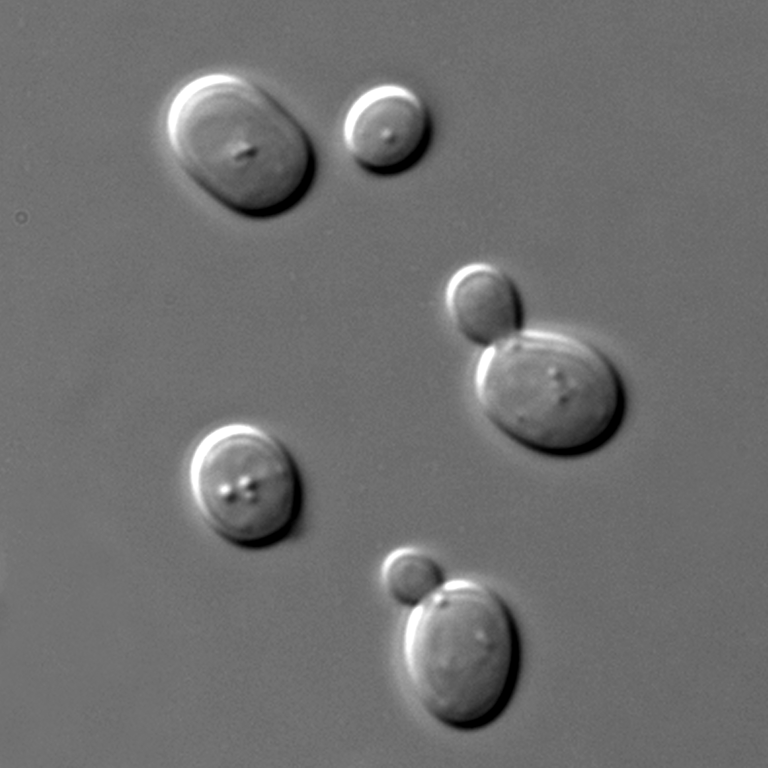
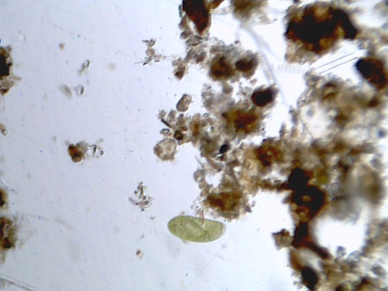
Download image provenance, source links and SHA-256 hashes. The original source files can be opened separately; no collection, culturing or unknown-organism handling is needed.
Reconstruct the original packet
The original schematic has 5 unambiguous connected objects but 7 unambiguous bodies. Including the faint feature gives 6 or 8 depending on the declared counting rule. Neither answer establishes viability.
Predict and investigate
Predict first: Can two defensible counting rules give different totals for the same attached objects?
Inspect both actual images and retain two visible features, one unresolved claim and exact source identity. Then compare both counting rules on the separate original schematic, including and excluding the ambiguous feature. Explain why neither actual species nor viable count follows from that schematic.
Defend a changed case
Apply the mechanism to a changed boundary
An analyst reports 8 after counting resolved bodies and including the faint circle. What would the connected-object rule excluding that feature report, and why is the difference not evidence of cell death?
Retain the first response, units, uncertainty and any prior feedback exposure in the notebook before comparing this public explanation.
Compare a worked response
It reports 5. Two attached pairs each change from two bodies to one connected object, and the uncertain feature is omitted:8−2−1=5. Only the analysis rule changed; no temporal or viability observation was made.
Retain evidence without overclaiming
Preserve the first response, original source identity, object definition, units, uncertainty and feedback exposure in the Microbiology course evidence workspace. Actual source-labeled images are separate from original diagrams and numerical packets. No sample collection, culturing, staining procedure or participant testing is requested. Independent unfamiliar reasoning and practical competence require separate assessor evidence.
Primary mechanism reference, checked 30 October 2026. Original prose, models and exercises; actual image reuse is documented separately.
Apply
Public practice checks a specific claim. It does not establish independent mastery.
An analyst reports 8 after counting resolved bodies and including the faint circle. What would the connected-object rule excluding that feature report, and why is the difference not evidence of cell death?
Continue
Completion requires a saved prediction, at least one capture and a reflection. Presence checks do not grade your reasoning.
Skipping retains experiments and awards no mastery.
Save a science backup or import one
Science work is stored locally in this browser, separately from mathematics and AI. It does not sync automatically. Use this science-only backup; this checkout's older all-subject export does not include science.
A legacy file is available only when the complete snapshot fits the 36 MB limit. For larger snapshots, prepare a complete file set below; never discard notebooks to make a smaller file.
Matching imported notebook drafts replace their current drafts. Omitted lessons and known first attempts remain. Export your current work first if you want to retain both drafts.
Complete file sets and course backups
A file set can hold more work than one science file. Keep its manifest and every part together. Each file is limited to 36 MB. This format needs this version of LearnAI or a compatible newer version; older versions can use only the legacy single-file backup above.
The set includes every saved science course and global continuation at this moment. Later edits need a new export. A conflicting tab exports its own visible work, which may differ from saved work in another tab.
0 file(s) selected. File order and filenames do not determine identity. Missing, changed or conflicting parts block the entire restore; no good part is applied by itself.
Science storage: loading. No account or remote computation is required.